
Wêne:Development of the neural tube.png
Versyonekî jê mezintir tune.
Development_of_the_neural_tube.png (598 × 368 pixel, mezinbûnê data: 36 KB, MIME-typ: image/png)
| Ev pel ji Wikimedia Commonsê ye. Agahdariya li ser rûpela danasîna pelê li jêr tê nîşandan. |
Danasîn
| Danasîn |
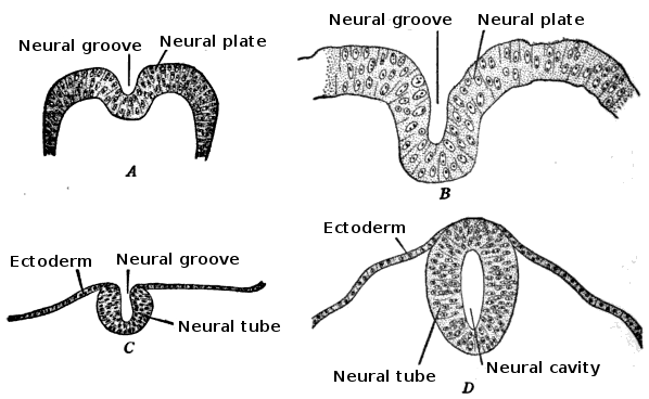
English: Development of the neural tube in human embryos (Prentiss-Arey). A. An early embryo (Keibel) B. at 2 mm. (Graf Spee) C. at 2 mm. (Mall) D. at 2.7 mm (Kollmann).
This is a scan of Figure 6 of the book "The anatomy of the nervous system" by Stephen Walter Ranson, with the labels redrawn. |
||||||||||||||||
| Dîrok | file created 2009-12-24, original image published 1920 | ||||||||||||||||
| Çavkanî |
Figure 6 (p. 24) of "The anatomy of the nervous system" by Stephen Walter Ranson, published W.B. Saunders, 1920
|
||||||||||||||||
| Xwedî | user:Looie496 created file, original artist unknown | ||||||||||||||||
| Guhartoyên din |
|
||||||||||||||||

== Lîsans ==
This media file is in the public domain in the United States. This applies to U.S. works where the copyright has expired, often because its first publication occurred prior to January 1, 1929, and if not then due to lack of notice or renewal. See this page for further explanation.
|
 | |
 |
This image might not be in the public domain outside of the United States; this especially applies in the countries and areas that do not apply the rule of the shorter term for US works, such as Canada, Mainland China (not Hong Kong or Macao), Germany, Mexico, and Switzerland. The creator and year of publication are essential information and must be provided. See Wikipedia:Public domain and Wikipedia:Copyrights for more details.
|
Dîroka daneyê
Ji bo dîtina guhartoya wê demê bişkoka dîrokê bitikîne.
| Dîrok/Katjimêr | Wêneyê biçûk | Mezinahî | Bikarhêner | Şirove | |
|---|---|---|---|---|---|
| niha | 20:08, 5 kanûna paşîn 2010 | | 598 x 368 (36 KB) | Looie496 | {{Information |Description={{en|1=Development of the neural tube in human embryos (Prentiss-Arey). A. An early embryo (Keibel) B. at 2 mm. (Graf Spee) C. at 2 mm. (Mall) D. at 2.7 mm (Kollmann). This is a scan of Figure 6 of the book "The anatomy of |
Bikaranîna pelê
Rûpelekî ku ji vê dosyeyê bi kar tîne nîne.
Bikaranîna gerdûnî ya pelê
Ev wîkiyên di rêzê de vê pelê bi kar tînin:
- Bikaranîna di af.wikipedia.org de
- Bikaranîna di ar.wikipedia.org de
- Bikaranîna di az.wikipedia.org de
- Bikaranîna di bg.wikipedia.org de
- Bikaranîna di bs.wikipedia.org de
- Bikaranîna di en.wikipedia.org de
- Bikaranîna di fr.wikipedia.org de
- Bikaranîna di gl.wikipedia.org de
- Bikaranîna di hr.wikipedia.org de
- Bikaranîna di id.wikipedia.org de
- Bikaranîna di sr.wikipedia.org de